UAH researchers discover ultrasound-based approach that may help reduce harmful inflammation and support joint healing
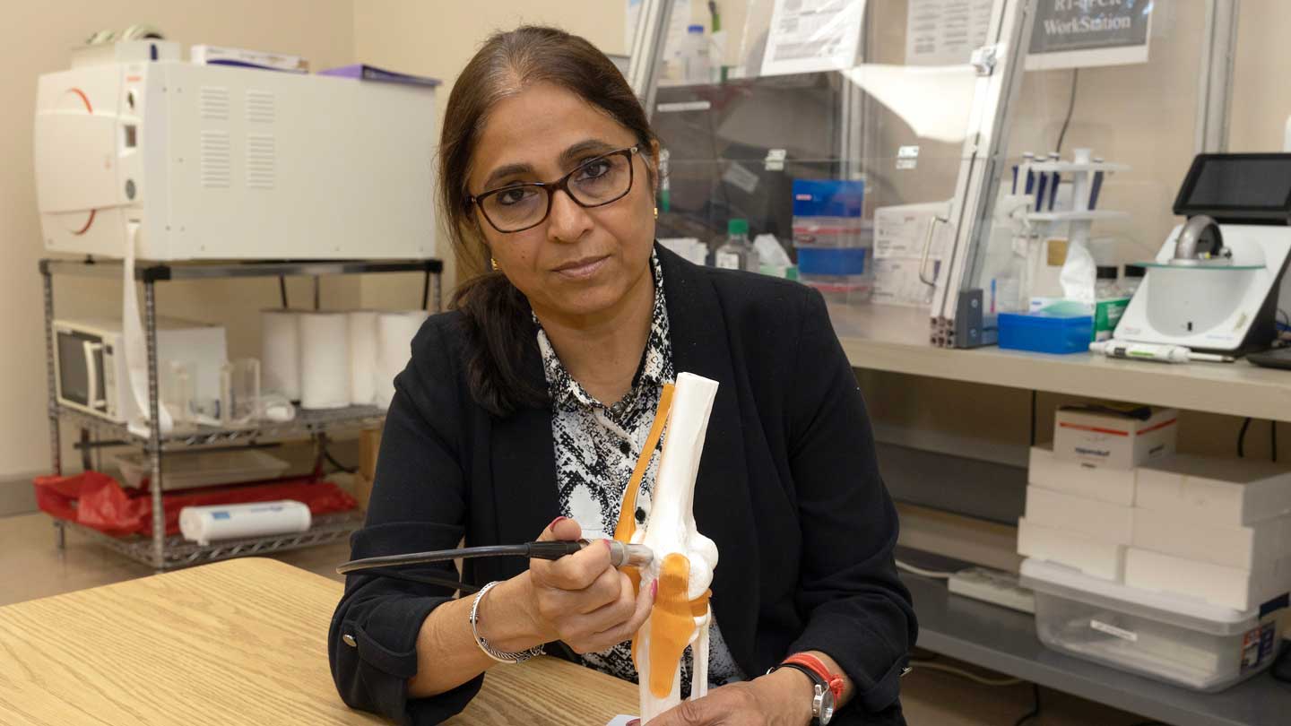
Dr. Anuradha Subramanian, professor of chemical and materials engineering.
With an aging population experiencing joint pain and inflammation at an all-time high, researchers at The University of Alabama in Huntsville (UAH), a part of The University of Alabama System, have published new findings that suggest continuous low-intensity ultrasound may help shift the body's immune response from prolonged inflammation toward tissue repair, a discovery that could eventually contribute to novel treatments for joint injuries and post-traumatic osteoarthritis.
The study, published in the Nature journal Scientific Reports, was conducted by a multidisciplinary team of UAH researchers under the leadership of Dr. Anuradha Subramanian, professor of chemical and materials engineering. The work brought together biological experimentation conducted by Dr. Shahid Khan as part of his doctoral work with computational and statistical methods developed by Dr. Satyaki Roy, professor of mathematical sciences, along with additional contributions from graduate student Owen Trippany. The research was supported by funding from the National Institutes of Health through an R01 grant awarded to Dr. Anuradha Subramanian.
The research examines how a non-invasive form of ultrasound affects macrophages, specialized immune cells that play a central role in both inflammation and healing.
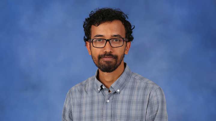
Dr. Satyaki Roy, professor of mathematical sciences.
“Following injury, the body recruits inflammatory 'defender' macrophages (M1) to clear damaged tissue and healer macrophages (M2) to support repair and recovery,” Subramanian explains. “Persistent dominance of defender macrophages can create a prolonged inflammatory environment that contributes to post-traumatic osteoarthritis.”
The UAH team investigated whether continuous low-intensity ultrasound could encourage macrophages to move away from this prolonged inflammatory state and toward one associated with tissue repair.
“In an ‘M1’ state, microphages promote inflammation to fight damage or infection, but prolonged M1 activity can also harm healthy tissue,” Subramanian notes. “In contrast, ‘M2-like’ macrophages support tissue repair and recovery. Shifting macrophages toward an M2-like state is important, because it may help reduce chronic inflammation while encouraging healing in damaged joints. Our findings suggest that continuous low-intensity ultrasound may help restore this balance by promoting a more reparative macrophage response."
“Post-traumatic osteoarthritis is driven in part by persistent inflammation that limits tissue repair and accelerates joint degeneration,” Roy adds. “Our team is interested in continuous low-intensity ultrasound because it offers a non-pharmacological, non-invasive approach that may help regulate immune cell behavior and promote a more reparative healing environment in injured joints.”
To better replicate what happens after a joint injury, the researchers used fibronectin fragments – molecules produced during tissue breakdown – rather than relying solely on traditional laboratory methods used to trigger inflammation. This innovation allowed the team to create a model that more closely resembles the biological environment inside an injured joint.
To support the study, Roy combined “transcriptomics,” the large-scale analysis of gene activity, with an advanced computational technique termed as “differential clustering.” This technique provides a way to find groups of genes that behave similarly when something changes, rather than just grouping genes that look similar in general. Instead of examining individual genes in isolation, the approach allowed researchers to identify coordinated changes across groups of genes, providing a broader view of how immune cells respond to ultrasound treatment.
"This allowed us to study not only which genes changed, but also how groups of genes changed their coordinated behavior in response to ultrasound stimulation," Roy says.
The results showed that continuous low-intensity ultrasound reduced markers associated with inflammation while increasing markers linked to a more reparative, M2-like macrophage state.
While the work remains at the laboratory research stage, the findings highlight the potential of non-drug, non-invasive technologies to influence immune behavior and support healing after injury. The researchers believe the approach could ultimately complement future therapies aimed at slowing the progression of osteoarthritis and improving recovery following joint trauma.
"The next steps will involve validating these findings in animal models of early post-traumatic osteoarthritis and studying how ultrasound-based modulation affects long-term tissue repair in joint injury settings,” Subramanian says.